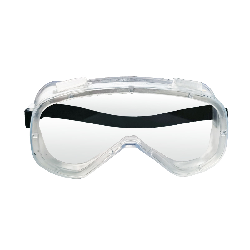
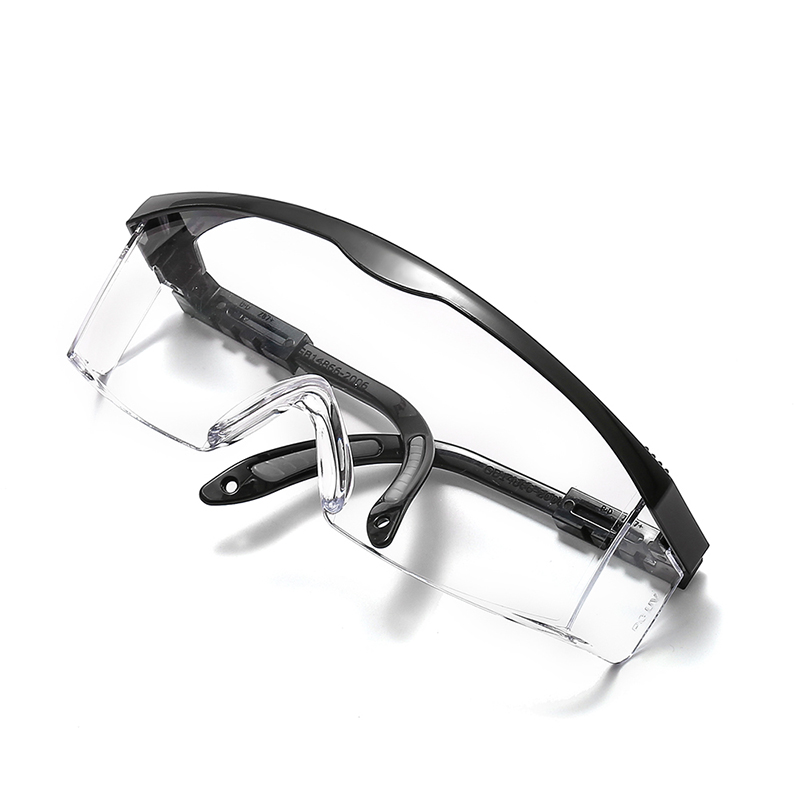
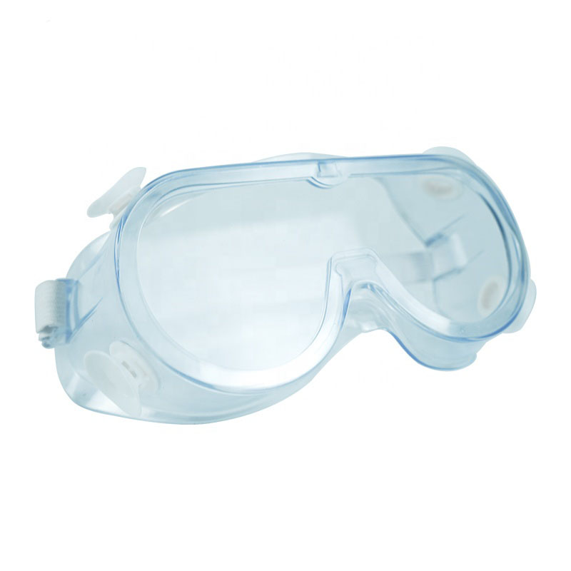
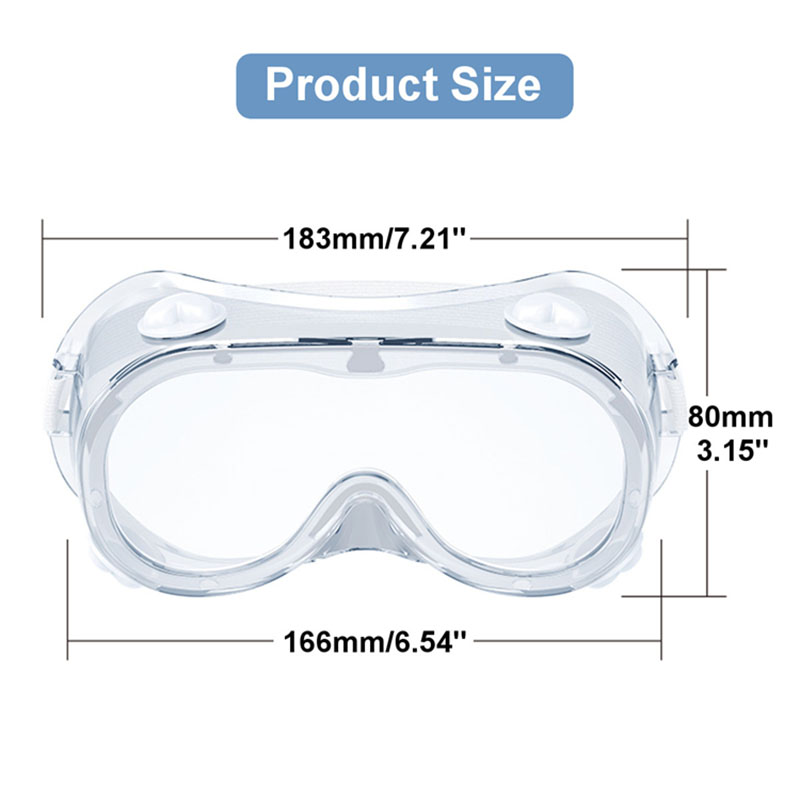
It can prevent some medicine or blood from splashing on the face, thus protecting the eyes. This kind of glasses is generally used in conjunction with masks and surgical caps to fully protect the doctor's head.
4. The breathability must be good, generally there are vents
Protective goggles,disposable medical equipment,personal protection,daily protection Shanghai Rocatti Biotechnology Co.,Ltd , https://www.ljdmedicals.com
[Functions of medical eye masks]
1. Prevent blood, potion and other liquids that cause damage to the skin from causing unpredictable damage to the eyes.
2. It can prevent the impact of objects on the eyes during close surgery.
3. The inner space of the eye mask is large enough for doctors who wear myopia glasses.


Source and solution for counting errors in blood cell counting boards - automated counting
introduction
The blood cell counting board has always been the gold standard for laboratory cell counting. Since the first use of blood samples from patients in the 18th century in France, blood cell counting plates have received a number of significant developments over the past few hundred years, which are more accurate, simpler to use, and ultimately formed. What we used today. The blood count plate count is still an integral part of all cytological studies, however the problems with its counting have not disappeared over time due to its inherent design and use. Here we will list the sources of errors in the counting of the blood cell counters and discuss how automated counting can eliminate these problems.
Source of counting error of blood cell counting board
1. Manual errors ( mixing, loading, dilution, calculation errors, and manual operation errors )
a. In the observation of 5 operators, the operation error and random error accounted for 3.12% and 7.8%, respectively [3] .
b. James M. Ramsey conducted an experiment to measure how the sampling area and dilution factor affect the accuracy of the count.
He tested three sampling areas (18, 9 and 4 mm 2 ) and two dilution factors (1:100 and 1:25). The CVs value is increased when the sampling area is reduced, and the increase in the dilution factor reduces the CVs [4] .
c. Bane found that when the same operator counted the same two semen samples, the difference in count results was 55% due to sampling and pipetting problems, and 45% due to counting chamber and cell counting problems [5] . Another experiment by Freund and Carol showed that the difference in counts between different operators can be as high as 52%, while the difference between the same operator is 20% [5] .
2. The need to count multiple times to ensure the accuracy of the results
a. In 1907, John C. DaCosta stated that in order to obtain accurate counting results, it is necessary to take multiple drops of blood from the blood sample for counting [1] .
b. Nielsen, Smyth and Greenfield concluded that in order to obtain 10%, 15% and 20% blood count plate count accuracy, the required number of samples were 7 copies, 3 copies and 2 copies, respectively. Contains 180, 200 and 125 cells [6] .
c. In 1881, Lyon and Thoma speculated that the standard error of the hemocytometer was, where n is the number of cells counted;
d. In 1907, William Sealy published the work of counting the brewer's yeast in the name of “studentâ€. He calculated the counting error through experiments and mathematical models. The formula is also [7,8] .
3. Requirements for uniform cell distribution
a. In 1912, James C. Todd listed the uneven distribution of cells as a source of error in counting errors [1] .
b. Students also stated that there are two main sources of counting error, one is that the extracted yeast sample cannot represent the concentration of the stock solution, and the other is that the cells are unevenly distributed in the counting area when randomly sampled [7,8] .
c. In 1947, an article referred to the problem of uneven distribution of cell concentrations in the hemocytometer. Initial results showed that the concentrations in the nearest and farthest regions from the inlet were 3.5% lower and 3.5% higher, respectively, than the average concentration [9] .
4. Instrument and material differences ( grid, depth, coverslip, buffer type and pipette )
a. The results show that the counting error of the counting chamber and the counting error caused by the pipette (CV%) are about 4.6% and 4.7%, respectively [10] .
b. In a counting experiment of 5 counting personnel, the errors caused by pipettes and hemocytometer were 9.46% and 4.26%, respectively [3] .
c. In 1961, Sanders and Skerry concluded that the position of the coverslips can cause a 7.6% difference in counts [11] .
d. In the counting experiments for different dilution steps, as the dilution step increases, the coefficient of variation increases and the error for each blood cell counting system is as follows: Bürker-Türk (BT) (7.7%-12%), Thoma (6.6 %-14.1%), Makler (19.8%-23.6%) [12] .
Solve the problem of counting the blood count board
With the development of new technologies, such as computer technology, automation software, optical lenses, fluorescent dyes, precision manufacturing, and modern technologies such as fluorescence microscopy, flow cytometry, image cytometry, automation has solved the existence of blood cell counting plates. Many questions [13-25] .
Automated counting solution:
Manual operation error - To solve this problem, automation and robotics can replace manual sample manipulation and counting operations. .
Loading error - The more sampling areas, the more cells count, the smaller the random error, but the more time it takes. By applying automated sampling or imaging techniques, millions of cells can be analyzed in a very short time, increasing efficiency and minimizing random errors in the analysis.
Pipetting and dilution error - These depend on the operator's operating experience. This error can be minimized by using an autosampler or an automatic flow system [26] .
Material error - The error in the counting chamber is due to differences between different brands of blood cell counting plates or different batches of the same brand. This can also be done through an automated cell counter (selection of the cell counter, please refer to http://dakewe.com/product/view/id-71.html or Baidu Library http://wenku.baidu.com/view/f13faf4916fc700abb68fc54 The article "Cell Counter Selection" in .html) increases the sampling amount and reduces the random error to solve.
Uneven cell distribution – Improper cleaning of the hemocytometer or incorrect placement of the coverslip will result in errors. These can be eliminated by a cell counter that does not use a counting chamber, such as a flow cytometer. However, if there are cell clusters in the cell sample, the instrument based on the flow count will be difficult to count, and using the image counter, the cell mass can use the image analysis algorithm to count the aggregated cells, which can improve the accuracy of the cell count.
Review
The blood cell counting board has been an indispensable tool in biomedical research for centuries, and has undergone many improvements to form what researchers use today, but it still causes many unavoidable counting errors. Today, the use of modern automated cell counters has largely eliminated many sources of error, increasing the accuracy and efficiency of cell counting.
Reference literature
1. Davis JD. THE HEMOCYTOMETER AND ITS IMPACT ON PROGRESSIVE-ERA MEDICINE. Urbana: University of Illinois at Urbana-Champaign; 1995.
2. Verso ML. Some Nineteenth-Century Pioneers of Haematology. Medical History 1971; 15(1): 55-67.
3. Biggs R, Macmillan RL. The Errors of Some Haematological Methods as They Are Used in a Routine Laboratory. Journal of Clinical Pathology 1948; 1: 269-87.
4. Ramsey JM. The Effects of Size of Sampling Area and Dilution on Leucocyte Counts in a Hemocytometer. The Ohio Journal of Science 1969; 69(2): 101-4.
5. Freund M, Carol B. Factors Affecting Haemocytometer Counts of Sperm Concentration in Human Semen. Journal of Reproductive Fertility 1964; 8: 149-55.
6. Nielsen LK, Smyth GK, Greenfield PF. Hemacytometer Cell Count Distribution: Implications of Non-Poisson Behavior. Biotechnology Progress 1991; 7: 560-3.
7. Student. On the Error of Counting with a Haemacytometer. Biometrika 1907; 5(3): 351-60.
8. Shapiro HM. "Cellular Astronomy" - A Foreseeable Future in Cytometry. Cytometry Part A 2004; 60A: 115-24.
9. Hynes M. The Distribution of Leucocytes on the Counting Chamber. Journal of Clinical Pathology 1947; 1: 25-9.
10. Berkson J, Magath TB, Hurn M. The Error of Estimate of the Blood Cell Count as Made with the Hemocytometer. American Journal of Physiology 1940; 128: 309-23.
11. Sanders C, Skerry DW. The Distribution of Blood Cells on Haemacytometer Counting Chambers with Special Reference to the Amended British Standards Specification 748 (1958). Journal of Clinical Pathology 1961; 14: 298-304.
12. Christensen P, Stryhn H, Hansen C. Discrepancies in the Determination of Sperm Concentration using Bürker-Türk, Thoma and Makler Counting Chambers. Theriogenology 2005; 63: 992-1003.
13. Al-Rubeai M, Welzenbach K, Lloyd DR, Emery AN. A Rapid Method for Evaluation of Cell Number and Viability by Flow Cytometry. Cytotechnology 1997; 24: 161-8.
14. Fazal SS. A test for a Generalized Poisson Distribution. Biometrical Journal 1977; 19(4): 245-51.
15. Hansen C, Vermeiden T, Vermeiden JPW, Simmet C, Day BC, Feitsma H. ​​Comparison of FACSCount AF system, improved neubauer hemocytometer, Corning 254 photometer, SpermVision, UltiMate and NucleoCounter SP-100 for determination of sperm concentration of boar semen Theriogenology 2006; 66(9): 2188-94.
16. Despotis GJ, Saleem R, Bigham M, Barnes P. Clinical evaluation of a new, point-of-care hemocytometer. Critical Care Medicine 2000; 28(4): 1185-90.
17. Paulenz H, Grevle IS, Tverdal A, Hofmo PO, Berg KA. Precision of the Coulter(R) Counter for Routine Assessment of Boar-Sperm Concentration in Comparison with the Hemocytometer and Spectrophotometer. Reproduction in Domestic Animals 1995; 30(3 ): 107-11.
18. Lutz P, Dzik WH. Large-Volume Hemocytometer Chamber for Accurate Counting of White Cells (Wbcs) in Wbc-Reduced Platelets - Validation and Application for Quality-Control of Wbc-Reduced Platelets Prepared by Apheresis and Filtration. Transfusion 1993; 33 (5): 409-12.
19. Brecher ME, Harbaugh CA, Pineda AA. Accurate Counting of Low Numbers of Leukocytes - Use of Flow-Cytometry and a Manual Low-Count Chamber. American Journal of Clinical Pathology 1992; 97(6): 872-5.
20. Vachula M, Simpson SJ, Martinson JA, et al. A Flow Cytometric Method for Counting Very Low-Levels of White Cells in Blood and Blood Components. Transfusion 1993; 33(3): 262-7.
21. Szabo SE, Monroe SL, Fiorino S, Bitzan J, Loper K. Evaluation of an Automated Instrument for Viability and Concentration Measurements of Cryopreserved Hematopoietic Cells. Laboratory Hematology 2004; 10: 109-11.
22. Chan LL, Wilkinson AR, Paradis BD, Lai N. Rapid Image-based Cytometry for Comparison of Fluorescent Viability Staining Methods. Journal of Fluorescence 2012; 22: 1301-11.
23. Chan LL, Zhong X, Qiu J, Li PY, Lin B. Cellometer Vision as an alternative to flow cytometry for cell cycle analysis, mitochondrial potential, and immunophenotyping. Cytom Part A 2011; 79A(7): 507-17.
24. Chan LL-Y, Lai N, Wang E, Smith T, Yang X, Lin B. A rapid detection method for apoptosis and necrosis measurement using the Cellometer imaging cytometry. Apoptosis 2011; 16(12): 1295-303.
25. Bocker W, Gantenberg HW, Muller WU, Streffer C. Automated cell cycle analysis with fluorescence microscopy and image analysis. Physics in Medicine and Biology 1996; 41(3): 523-37.
26. Macfarlane RG, Payne AM-M, Poole JCF, Tomlinson AH, Wolff HS. An Automatic Apparatus for Counting Red Blood Cells. British Journal of Haemacytology 1959; 5: 1-15.
Contributed by: Dako for Biotechnology Co., Ltd.